- Visibility 55 Views
- Downloads 16 Downloads
- DOI 10.18231/j.sajhp.2022.019
-
CrossMark
- Citation
Role of computed tomography (CT) scan in patients with blunt chest trauma
- Author Details:
-
Nirmala C. Chudasama
-
Darshan M. Patel *
-
Divangi P. Patel
-
Valay H. Shah
Introduction
Chest trauma is classified as blunt or penetrating, with blunt trauma being the cause of most thoracic injuries. The main difference in penetrating trauma is opening the thoracic cavity, created either by stabbing or gunshot wounds, which is absent in blunt chest trauma.[1] Following head and extremities injuries, Blunt thoracic injuries are the third most common injury in polytrauma patients.[2] Generally, blunt chest trauma is directly responsible for 27% of all trauma deaths and is a major contributor in another 55% of trauma-related deaths.
Several studies have shown that CT scanning is accurate in visualising intrathoracic injuries, such as Pneumothorax, haemothorax and lung contusions. In addition the availability, reliability and low complication rate of CT scans has lead to its widespread use in the evaluation of blunt trauma. [3]
Aims & Objectives
Assessment of blunt chest trauma patients with radiographic finding suggestive of pneumothorax, rib fractures, effusion and suspected lung injuries.
Materials and Methods
A retrospective review of patients with blunt chest trauma who were treated in the trauma centre and who had received chest CT scan. Data collection extracted from medical records.
A cases of 100 patients were included in the study with the age group between 18-49 years and out of 100 patients, 80 were males and 20 were females with mean age group 42.5 year. Patients referred from the trauma centre with blunt chest trauma due to road traffic accidents and high velocity trauma. The hospital staff collects data of all trauma patients who were transferred-into the trauma centre with blunt chest trauma.
Results
The most common mode of injury in blunt thoracic trauma was motor vehicle accidents. 60 patients presented within six hours of injury, 20 within 6-12 hours, 15 within 12-24 hours and remaining 5 after 24 hours.
45 patients underwent CT within 2-6 hours, 25 within 6-12 hours, 20 within 1-7 days and 10 after seven days. Various signs and symptoms were chest pain or tenderness, respiratory distress, surgical emphysema, decreased air entry, vomiting and abdominal guarding.
Pneumothorax was detected in 28 patients ([Figure 1]: a and b), 19 patients had associated rib fracture and 11 patients had associated pleural effusion. 11 patients were associated with subcutaneous emphysema ([Figure 1]c). In 19 patients, underlying lung parenchymal injury was present.

Hemothorax was detected in 15 patients on CT scan ([Figure 2]). Pneumomediastinum was detected in 3 patients ([Figure 3]), while pneumopericardium was not seen in any case.
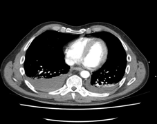
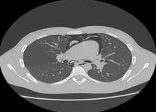
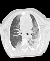
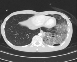
Pulmonary contusion was detected in 15 patients ([Figure 4]). Out of 100 patients of blunt thoracic trauma, pulmonary laceration was detected in 3 patients ([Figure 5]). Rib fracture was detected on CT scan in 53 patients ([Figure 6]).
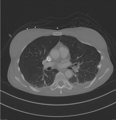
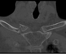
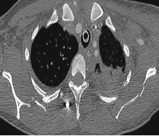
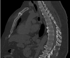
Clavicle fractures were detected in 11 patients on CT scan ([Figure 7]). Scapular fractures were detected in 7 patients on CT scan ([Figure 8]). Sternal fractures were detected in 6 patients on CT ([Figure 9]).
Discussion
In this study, CT chest scanning was significantly more effective in detecting Pneumothorax and haemopneumothorax, lung contusions and mediastinal haematomas. CT chest scan was also significantly better at detecting fractured ribs, scapulas, sternums and vertebrae.
Rib fracture is the most common injury in blunt chest trauma, and CT is the best method of visualization. [4] Rib fractures have been detected in half of the patients with blunt chest trauma. [5], [6]
Rib fracture occur in 56% of patients with major blunt chest trauma.10 Fractures of the lower ribs should increase the suspicion for upper abdominal organ. [7]
Scapular fracture is a rare finding in patients with chest trauma and has been reported in 3.7%–7.9% of those with blunt chest trauma. [8]
With the introduction of seat belt of legislation there is rise in sternal fracture.[9] Fractures of sternum are frequent, occurring in 1.5% to 4% of blunt chest trauma.
Clavicle fractures are usually obvious on the clinical examination. The most important role of MDCT in clavicle fracture evaluation relies in the assessment of medial fractures and injuries affecting the sternoclavicular joint, especially in the diagnosis of sternoclavicular dislocation. [10] Clavicular fractures from blunt chest trauma accounts for 3% of all dislocations.
Fractures involving the thoracic outlet, upper ribs, sternum, scapula and clavicle are significant because they are accompanied by brachial plexus/vascular injury in 3% to 15% of patients.
|
Abnormality |
No. of Abnormalities |
|
Pneumothorax |
28 |
|
Hemothorax |
15 |
|
Pneumomediastinum |
3 |
|
Subcutaneous emphysema |
11 |
|
Lung Contusion |
15 |
|
Lung Laceration |
3 |
|
Rib Fractures |
53 |
|
Clavicle Fractures |
11 |
|
Scapula Fractures |
7 |
|
Sternal Fractures |
6 |
Pneumothorax, an abnormal collection of air in the pleural space, is commonly seen after both blunt and penetrating trauma. An open wound may result in air entering the pleural space from the outside environment (an open pneumothorax or sucking chest wound), whereas in closed trauma, a pneumothorax is the result of lung parenchymal or airway injury that allows air to leak into the pleural space. [11]
Pneumomedistinum represents extra alveolar air in the mediastinum, a consequence of both blunt and penetrating trauma. Computed tomography imaging is much more sensitive for detection of pneumomediastinum particularly adjacent to the central pulmonary arteries, and aorta.[12]
Subcutaneous emphysema can occur in critically ill patients after blunt trauma to the chest and result in a pressure gradient between the intra-alveolar and perivascular interstitial space.[13]
Pulmonary contusions are the most common type of pulmonary parenchymal injury, occurring in up to 75% of blunt chest trauma cases.[14]
Pulmonary laceration occurs in major chest trauma when disruption and tearing of the lung parenchyma follows shearing forces, caused by direct impact, compression or inertial deceleration. [15]
Conclusion
Computed Tomography(CT) Scan is the modality of choice for rapid assessment of emergency chest trauma patients where associated abdominal injuries can be scanned in one sitting with high sensitivity and specificity.
Conflict of Interest
None.
Source of Funding
None.
References
- K Shanmuganathan, J Matsumoto. Imaging of penetrating chest trauma. Radiol Clin North Am 2006. [Google Scholar] [Crossref]
- R Kaewlai, LL Avery, AV Asrani, RA Novelline. Multidetector CT of blunt thoracic trauma. Radiographics 2008. [Google Scholar] [Crossref]
- S Peters, V Nicolas, CM Heyer. Multidetector computed tomography-spectrum of blunt chest wall and lung injuries in polytraumatized patients. Clin Radiol 2010. [Google Scholar] [Crossref]
- S Sridhar, C Raptis, S Bhalla. Imaging of blunt thoracic trauma. Semin Roentgenol 2016. [Google Scholar]
- A Ikonomou, P Prassopoulos. CT imaging of blunt chest trauma. Insights Imaging 2011. [Google Scholar]
- MA Afacan, F Buyukcam, UY Cavus. Acil Servise Başvuran Künt Toraks Travma Vakalarinin Incelen [The evaluation of patients with blunt thoracic trauma in the emergency room. Kocatepe Med J 2012. [Google Scholar]
- G P Sangster, A González-Beicos, A I Carbo. Blunt traumatic injuries of the lung parenchyma, pleura, thoracic wall, and intrathoracic airways: multidetector computer tomography imaging findings. Emerg Radiol 2007. [Google Scholar] [Crossref]
- S Wirth, S Jahnsen, M Mariano Scagliono, U Linsenmaier, G Schueller, S Wirth. Bony and thoracic chest wall injuries. Bony and Thoracic Chest Wall Injuries 2017. [Google Scholar]
- J S Budd. Effect of seat belt legislation on the inci-dence of sternal fractures seen in the accident depart-ment. Br Med J (Clin Res Ed) 1985. [Google Scholar] [Crossref]
- H Mirka, J Ferda, J Baxa. Multidetector computed tomography of chest trauma: indications, technique and interpretation. Insights Imaging 2012. [Google Scholar] [Crossref]
- . Chest radiography in thoracic polytrauma. AJR Am J Roentgenol 2009. [Google Scholar]
- M Wintermark, P Schnyder. The Macklin effect: a frequent etiology for pneumomediastinum in severe blunt chest trauma. Chest 2001. [Google Scholar] [Crossref]
- RJ Maunder, DJ Pierson, LD Hudson. Subcutaneous emphysema. Pathophysiology, diagnosis, and management. Arch Intern Med 1984. [Google Scholar]
- A Oikonomou, P Prassopoulos. P. CT imaging of blunt chest trauma. Insights Imaging 2011. [Google Scholar] [Crossref]
- RB Wagner, WO CrawfordJr, PP Schimpf. Classification of parenchymal injuries to the lung. Radiology 1988. [Google Scholar]
