- Visibility 106 Views
- Downloads 20 Downloads
- DOI 10.18231/j.sajhp.2023.016
-
CrossMark

Turkey ear as a cutaneous manifestation of tuberculosis
Introduction
Lupus vulgaris is the most common morphological variant of cutaneous tuberculosis. Cutaneous tuberculosis is rare today and is often confused with other granulomatous lesions. Its diagnosis remains bothersome because detecting mycobacteria in skin lesions using a conventional laboratory examination remains difficult. Turkey ear is a clinically descriptive term, previously being used for the earlobe with reddish indurated plaque lesions, which recently can be a sign for lupus vulgaris.
Case Report
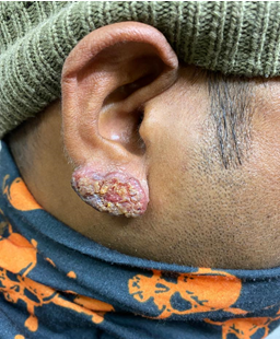
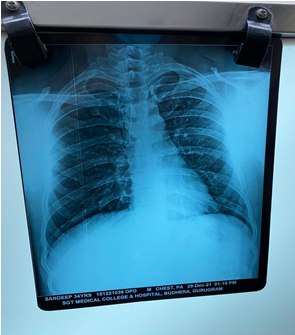
A 34-year-old man presented with an asymptomatic elevated skin lesion over the right ear for 6 months. The patient was apparently well 6 months back when he started developing swelling, the size of a mustard seed over the right ear. The patient then went to a local practitioner where local excision of the swelling was done. The wound wasn’t healed and it became a raw area and it further increased in size over a couple of days. The patient then visited the Ear, Nose, and Throat (ENT) Department at SGT Medical College and Gurugram, Haryana, India where a biopsy of the wound was done. There was no itching or any pain associated with the swelling. There was no lymphadenopathy. The patient had a history of ear piercing 20 days back. On examination, a single well-defined plaque is present over the lobe of the right ear of size 3*2 cm, erythematous and irregular surface with some crustation and oozing, non-tender and indurated lesion. There were no associated constitutional symptoms, history of previous trauma Bacille Calmette-Guerin (BCG) vaccination, or medical history of systemic tuberculosis. The rest of the systemic examination was within normal limits. Skin biopsy report shows stratified squamous epithelium with granulomatous lesion with dense lymphocytic infiltrate at the dermo-epidermal junction along with epithelioid cell granuloma and giant cells in the dermis. The patient was then referred to the dermatology department for further management. Routine blood investigations were done which revealed hemoglobin (Hb) 14.5 gm/dl, total leukocyte count (TLC) 7500/cmm3, and platelet count 1,50,000/mm3. Mantoux test was done which was positive. The diagnosis of plaque form of lupus vulgaris was depending on clinical and histopathological findings and anti-tubercular treatment of rifampicin, isoniazid, pyrazinamide, and ethambutol was started according to weight band for 6 months. The patient was then referred to the department of pulmonary medicine to rule out pulmonary tuberculosis. The patient chest x-ray (PA view) was done which was normal. The patient was advised sputum for acid-fast bacilli and GeneXpert and was advised follow-up after a period of 7 days.
Discussion
A very rare form of extrapulmonary tuberculosis is cutaneous tuberculosis[1] As over time resistance to the antitubercular drugs has been seen. Diseases and immunodeficiency syndromes such as AIDS and chemotherapy have caused tuberculosis to increase recently. So, this has also led to an increase in the incidence of cutaneous tuberculosis.[2] The most common morphological variant of cutaneous tuberculosis is lupus vulgaris. The average prevalence of cutaneous tuberculosis is 0.37% among general skin patients.[1] It is usually reinfection tuberculosis of the skin, and origination is from the focus of tuberculosis somewhere else in the body. It spreads by either hematogenous or lymphatic route or in a contagious way. It is rarely seen after BCG vaccination or it occurs after exogenous inoculation. However, the exact way in which LV develops is difficult to assess in many cases. It is more commonly seen in females as compared to males. It equally affects all age groups.[3] The classical presentation is a well-demarcated soft in consistency reddish-brown color papules or small plaques and is often located in the head and neck region. On diascopy the lesions which are deep-seated look like a yellowish-brown or "apple-jelly" color.[1], [2], [3], [4], [5], [6], [7] Conventional morphological patterns of LV are nodular, papular, ulcerative, vegetating, plaque, and tumid forms. Lichen simplex chronicus ulcerovegetating, frambesiform, gangrenous, sporotrichoid, myxomatous types are unusual variants.[1] There is large and huge soft tumors which occur mostly on the earlobes in the myxomatous form, which over time become grossly enlarged. The index of suspicion may be low because it is a rare form. [4] Over 80% of lesions are on the head and neck, particularly around the nose in Europe.[5], [8] LV lesions are mostly chronic, with new plaques appearing at the site of atrophic or regressed ones, and require proper treatment for complete cure and healing.[2] If not adequately treated, they can remain for many years and even decades. A combination of clinical, bacteriological and histological criteria is necessary to make the diagnosis of LV.[1], [2] LV is mostly seen in individuals with moderate immunity and a high amount of sensitive to tuberculin. Its diagnosis remains worry some, because mycobacteria is difficult to be detected in skin lesions using conventional laboratory methods and cultures are also often negative.[5] Diagnosis by both, detection of mycobacterial DNA using PCR and culture and has been reported and may be useful where mycobacteria are present in small numbers.[4] Histopathologically, typical necrotic caseating granulomatous tubercule with Langhans giant cells, mononuclear infiltrate and epitheloid cells can be seen.
The treatment of LV is by conventional antitubercular therapy (ATT) which consists of rifampicin, pyrazinamide, isoniazid, and ethambutol for 6 months. This should be given according to the weight band of the patient.[4], [6], [7] If LV is not properly treated, it presents as a progressive chronic development and its long- term complications can include cutaneous neoplasms.[9] Very often LV is confused with various cutaneous diseases and some other granulomatous processes of the skin as tuberculoid leprosy, cutaneous lymphoma, lupus pernio, deep-seated mycosis, lupoid leishmaniasis, and even granuloma annulare.[1], [4], [6], [7], [8] The most confusing diagnosis with earlobe lupus vulgaris is lupus pernio. Lupus pernio is a relatively common skin manifestation of sarcoidosis which is accompanied by slowly progressive violaceous indurated plaques and nodules which are bluish-red in colour and that usually affect the nose, cheeks, hands, fingers, ears, and toes. "Turkey ear" is a descriptive term previously used in lupus pernio of earlobe.[1] Williams et al. reported a case of lupus vulgaris, which had appearance of a turkey ear and suggested if turkey ear could be thought as a clinically descriptive term.[4]
In our case, localization and duration of the lesion were very typical for LV, in which the diagnosis was supported by tuberculin hypersensitivity and histopathological findings. The clinical appearance was very similar to turkey ear, which was previously described once as a sign of lupus vulgaris.[4] This is the third reported case of "turkey ear" as a manifestation of cutaneous tuberculosis. Thus, the term "turkey ear" is supposed to be used not only for sarcoidosis but also for lupus vulgaris.
Source of Funding
None.
Conflict of Interest
None.
References
- S Khandpur, BSN Reddy. Lupus vulgaris: unusual presentations over the face. J Eur Acad Dermatol Venereol 2003. [Google Scholar]
- IK Altunay, Z Baysal, TR Ekmekçi, A Köslü. Incidence of cutaneous tuberculosis in patients with organ tuberculosis. Int J Dermatol 2003. [Google Scholar]
- A Wozniacka, R Schwartz, AS Jedrzejowska, M Borun, C Arkuszewska. Lupus vulgaris: Report of two cases. Int J Dermatol 2005. [Google Scholar]
- C Williams, A Mitra, S Walton. Turkey ear': A diagnosis or a physical sign. Br J Dermatol 2007. [Google Scholar]
- M Okazaki, A Sakurai. Lupus vulgaris of the earlobe. Ann Plast Surg 1997. [Google Scholar]
- M Senol, A Ozcan, B Mizrak, AC Turgut, S Karaca, H Kocer. A case of lupus vulgaris with unusual location. J Dermatol 2003. [Google Scholar]
- E Ozkaya-Beyazit, C Baykal, N Buyukbabani, G Hafiz. Earlobe dermatitis. Arch Dermatol 2002. [Google Scholar]
- C Ceylan, B Gerceker, F Ozdemir, A Kazandi. Delayed diagnosis in a case of lupus vulgaris with unusual localization. J Dermatol 2004. [Google Scholar]
- TR Ekmekci, A Koslu, D Sakiz, M Ozcivan. Squamous cell carcinoma arising from lupus vulgaris. J Eur Acad Dermatol Venereol 2005. [Google Scholar]